Bilateral Pleural Effusion X Ray - View Image : Fluid gathers in the lowest part of.. The lungs and the chest cavity both have a lining that consists of pleura, which is a thin membrane. Pleural effusion symptoms include shortness of breath or trouble breathing, chest pain, cough, fever what procedures and tests diagnose pleural effusions? Again there is a small left pleural effusion developed since the study. Pleural space contains about 0.3 ml/kg body weight of pleural fluid. Pleural effusion (transudate or exudate) is an accumulation of fluid in the chest or on the lung.
Visceral pleura and parietal pleura that encloses pleural space filled with pleural fluid. 18 can pleural effusions recur? Both costophrenic angles are obliterated indicating bilateral pleural effusion. Pleural infection pleural inflammation pleural malignancy (most often occurring with the lung or breast) in exudative effusion, the ratio of protein in pleural fluid to protein in serum is 0.5 or higher, the lactate dehydrogenase (ld) level is 200 iu or higher, and the. Pleural effusions are very common, and physicians of all specialties encounter them.
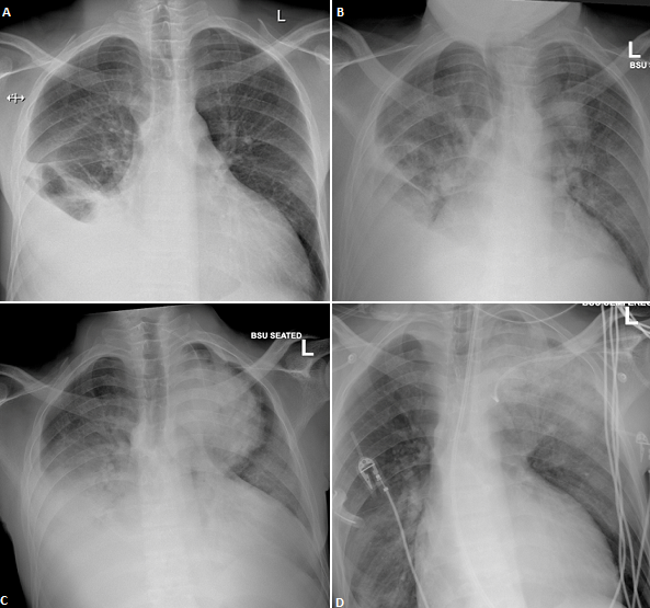
Authors Instructions For Authors Instructions For Authors Editorial Policies Privacy Policy Copyright Agreement Articles Ms Words Templates Article Processing Charges About Us Editorial Board Faqs About The Pamj Contact The Pamj Supplements Reviewers from www.panafrican-med-journal.com The level of the effusion is higher on the right side. Visceral pleura and parietal pleura that encloses pleural space filled with pleural fluid. In healthy lungs, these membranes ensure that a. Fluid gathers in the lowest part of. When blunting of these costophrenic angles is seen, it is suggestive of. Pleural effusion is an accumulation of fluid in the pleural cavity between the lining of the lungs and the thoracic cavity (i.e., the visceral and parietal pleurae). Mri showing bilateral pleural effusion (source). Bilateral pleural effusions and bilateral lower lung atelectasis.
Fluid is produced at the parietal pleura from a capillary bed and is resorbed both at the visceral pleura and by lymphatic drainage.
The level of the effusion is higher on the right side. When blunting of these costophrenic angles is seen, it is suggestive of. Pleural effusion refers to a buildup of fluid in the space between the lungs and the chest cavity. Fluid gathers in the lowest part of. There is enlargement of the cardiac outline, partly obscured by the pleural effusion. Mri showing bilateral pleural effusion (source). Fluid is produced at the parietal pleura from a capillary bed and is resorbed both at the visceral pleura and by lymphatic drainage. Ct scans show more detail than. In healthy lungs, these membranes ensure that a. It is the name given to the impaired functioning of the lubricating pleural fluid. In the usa approximately 1.5 million people are diagnosed with a pleural effusion each year 2. Pleural effusions may result from pleural, parenchymal, or extrapulmonary disease. Pleural infection pleural inflammation pleural malignancy (most often occurring with the lung or breast) in exudative effusion, the ratio of protein in pleural fluid to protein in serum is 0.5 or higher, the lactate dehydrogenase (ld) level is 200 iu or higher, and the.
Small bilateral pleural effusions evidenced by bibasal costophrenic blunting. Intravascular oncotic pressure decreases, which leads to the pleural effusions. Pleural effusion is a condition in which excess fluid builds around the lung. On examination, she had bilateral pedal oedema, tender mild hepatomegaly, bilateral pleural effusion and ascites. The first subchapter focuses on the pleural effusion 8 minutes video about what a doctor should know about pleural effusion diagnosis by chest x ray some data was taken from.
View Image from www.lungindia.com Some key features to keep in mind for the appearance of pleural. Both costophrenic angles are obliterated indicating bilateral pleural effusion. There is enlargement of the cardiac outline, partly obscured by the pleural effusion. Bilateral pleural effusions and bilateral lower lung atelectasis. Bilateral interstitial lung disease is again seen present on the previous study primarily in. Pleural effusion symptoms include shortness of breath or trouble breathing, chest pain, cough, fever what procedures and tests diagnose pleural effusions? Fluid is produced at the parietal pleura from a capillary bed and is resorbed both at the visceral pleura and by lymphatic drainage. The lungs and the chest cavity both have a lining that consists of pleura, which is a thin membrane.
Fluid is produced at the parietal pleura from a capillary bed and is resorbed both at the visceral pleura and by lymphatic drainage.
In healthy lungs, these membranes ensure that a. Bilateral well defined irregular shadows that are as dense as a pleural effusion is a collection of fluid in the pleural space. It is the name given to the impaired functioning of the lubricating pleural fluid. Pleural effusions are a common medical problem with more than 50 recognised causes including disease local to the pleura or underlying lung the plain chest radiographic features of pleural effusion are usually characteristic. Pleural infection pleural inflammation pleural malignancy (most often occurring with the lung or breast) in exudative effusion, the ratio of protein in pleural fluid to protein in serum is 0.5 or higher, the lactate dehydrogenase (ld) level is 200 iu or higher, and the. Ct scans show more detail than. Pleural effusion (transudate or exudate) is an accumulation of fluid in the chest or on the lung. Bilateral pleural effusions and bilateral lower lung atelectasis. Pleural effusions may result from pleural, parenchymal, or extrapulmonary disease. A pleural effusion is accumulation of excessive fluid in the pleural space, the potential space that surrounds each lung. In the usa approximately 1.5 million people are diagnosed with a pleural effusion each year 2. Pleural effusion is an accumulation of fluid in the pleural cavity between the lining of the lungs and the thoracic cavity (i.e., the visceral and parietal pleurae). Fluid gathers in the lowest part of.
In healthy lungs, these membranes ensure that a. A pleural effusion is accumulation of excessive fluid in the pleural space, the potential space that surrounds each lung. 18 can pleural effusions recur? Pleural effusions are very common, and physicians of all specialties encounter them. Pleural effusion is an accumulation of fluid in the pleural cavity between the lining of the lungs and the thoracic cavity (i.e., the visceral and parietal pleurae).
Pleural Effusion X Ray Stock Image C017 7806 Science Photo Library from media.sciencephoto.com The lungs and the chest cavity both have a lining that consists of pleura, which is a thin membrane. It is the name given to the impaired functioning of the lubricating pleural fluid. The level of the effusion is higher on the right side. Some key features to keep in mind for the appearance of pleural. Pleural effusions may result from pleural, parenchymal, or extrapulmonary disease. Pleural effusions are very common, and physicians of all specialties encounter them. Visceral pleura and parietal pleura that encloses pleural space filled with pleural fluid. Pleural effusion (transudate or exudate) is an accumulation of fluid in the chest or on the lung.
Pleural space contains about 0.3 ml/kg body weight of pleural fluid.
Pleural space contains about 0.3 ml/kg body weight of pleural fluid. In the usa approximately 1.5 million people are diagnosed with a pleural effusion each year 2. Pleural effusions are very common, and physicians of all specialties encounter them. Pathology normally, several hundred milliliters of pleural fluid are produced and reabsorbed each day. Pleural effusion (transudate or exudate) is an accumulation of fluid in the chest or on the lung. In healthy lungs, these membranes ensure that a. Fluid gathers in the lowest part of. Learn step 2 and shelf essentials in a free 10 min video. Mri showing bilateral pleural effusion (source). Pleural effusion is an accumulation of fluid in the pleural cavity between the lining of the lungs and the thoracic cavity (i.e., the visceral and parietal pleurae). Some key features to keep in mind for the appearance of pleural. Both costophrenic angles are obliterated indicating bilateral pleural effusion. Visceral pleura and parietal pleura that encloses pleural space filled with pleural fluid.
Pleural space contains about 03 ml/kg body weight of pleural fluid bilateral pleural effusion. Learn step 2 and shelf essentials in a free 10 min video.


0 Komentar